For the past 4 years, a 14-year-old girl from Ninh Binh had been living with epileptic seizures occurring many times a day, severely affecting her learning and daily life. After undergoing surgery at the Neurology Center to place cortical surface electrodes and deep electrodes, map the epileptogenic zone, and remove a complex seizure focus, the child is now free from the prolonged seizures that had affected her for years. This is one of the world’s advanced epilepsy surgery techniques that Vietnam National Children’s Hospital has mastered and successfully implemented for many years.
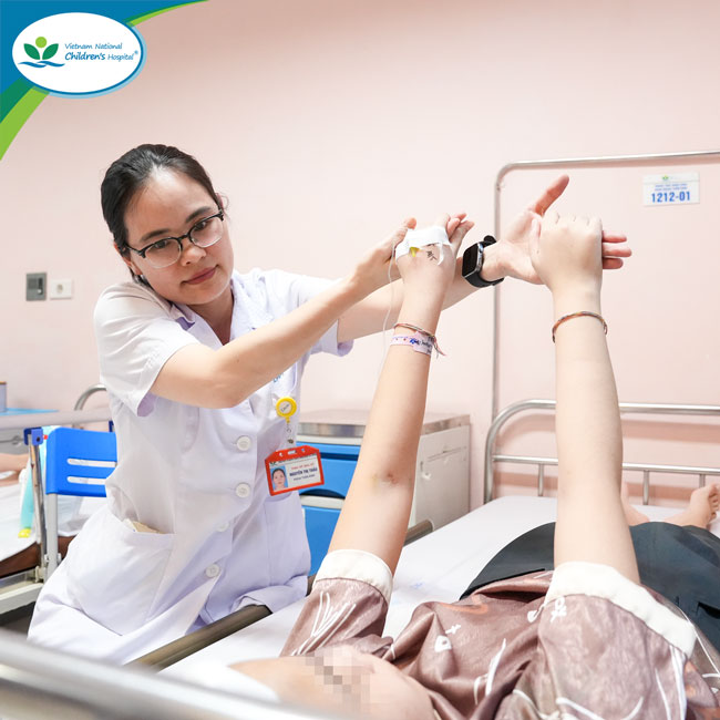
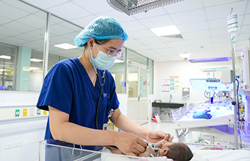
Dr. Nguyen Thi Thao, MSc, MD (Specialist Level II), examined and assessed the patient’s neurological function before discharge.
The journey to locate an “epileptogenic focus” with no lesion on MRI
For 4 years since being diagnosed with epilepsy in 2022, the 14-year-old girl had faced seizures every day. The seizures occurred suddenly and were especially easy to trigger by auditory stimuli such as a rooster crowing, a ringing phone, or vehicle horns.
Frequent seizures caused the child to fall many times, leading to injuries to the head and face. Beyond physical health, prolonged epilepsy also severely affected her cognitive development. From an ordinary student, she gradually struggled with learning, her ability to absorb knowledge declined, and eventually she could no longer continue attending school. Before surgery, her IQ had decreased to 70.
At Vietnam National Children’s Hospital, she was diagnosed with focal epilepsy and treated with five anti-seizure medications, but still had 2–10 seizures per day, and sometimes up to 30 seizures a day.
At the Neurology Center, the child underwent a specialized assessment to identify the epileptogenic focus for surgical treatment. However, brain MRI did not detect any lesion, creating a major challenge.
According to Dr. Nguyen Thi Thao, MSc, MD (Specialist Level II), Neurology Functional Exploration Unit, Neurology Center, Vietnam National Children’s Hospital, identifying the epileptogenic focus in children with epilepsy and no visible lesion on brain MRI is a major challenge—not only in Viet Nam but also at major epilepsy surgery centers worldwide.
After careful analysis of clinical findings, neurological examination, video-EEG, and PET imaging results, the epilepsy surgery team at Vietnam National Children’s Hospital decided to perform a two-stage surgery:
Stage 1: placement of cortical surface electrodes and deep EEG electrodes.
A total of 98 electrodes were placed on the brain surface and within brain tissue.
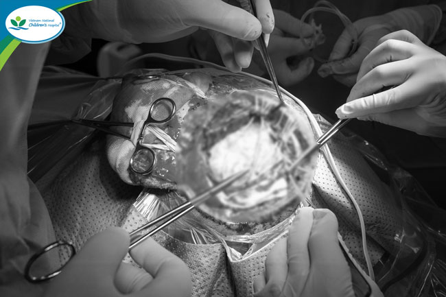
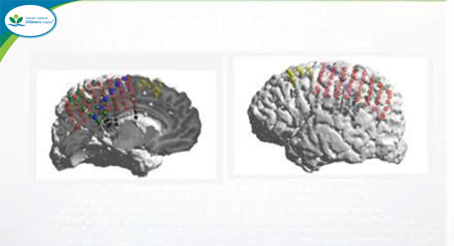
The intracranial electrode system was used to precisely identify the epileptogenic zone and map cortical function before surgery.
The patient was monitored and underwent stimulation testing at the Neurology Center to identify the epileptogenic zone while preserving motor function. This is a complex technique requiring a deep understanding of neuroanatomy and neurological function, seizure characteristics, and the relationship between clinical manifestations and EEG findings.
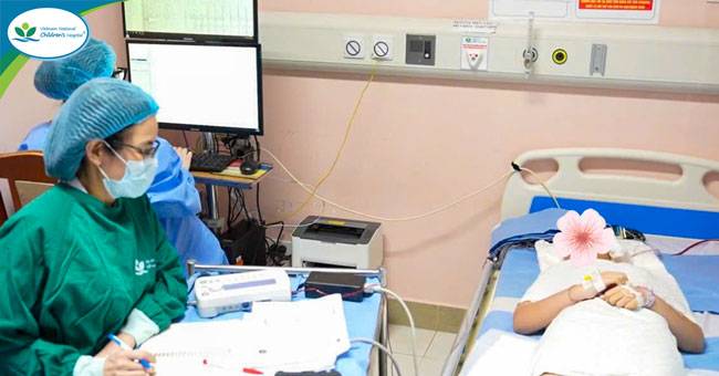
After 3 days of monitoring and identification of the epileptogenic zone, on 15/05/2026, the child was taken to the operating room for Stage 2 surgery: resection of the epileptogenic zone.
Ending 4 years of prolonged seizures through advanced epilepsy surgery
Dr. Le Nam Thang, MD (Specialist Level II), Head of the Division of Neurosurgery and Deputy Director of the Neurology Center, Vietnam National Children’s Hospital, explained that epilepsy surgery is a specialized treatment approach aimed at removing or isolating the brain region responsible for seizures.
Most children with drug-resistant epilepsy have underlying structural brain abnormalities, such as cortical dysplasia or early neurodevelopmental disorders. Although many anti-seizure medications are available, about 30–40% of patients do not respond to medical treatment. For these patients, surgery is considered an effective solution to control or stop seizures.

Dr. Le Nam Thang noted that epileptic seizures often last only a few seconds and can easily be missed if repeated unusual symptoms are not carefully noticed.
For complex epilepsy cases, surgery combined with cortical surface or intracranial electrode placement to map the epileptogenic zone requires close coordination among multiple specialties. From electrode placement and intraoperative EEG interpretation to anesthetic adjustment, every step must be performed precisely to accurately identify the seizure-generating area for removal while ensuring maximum patient safety.
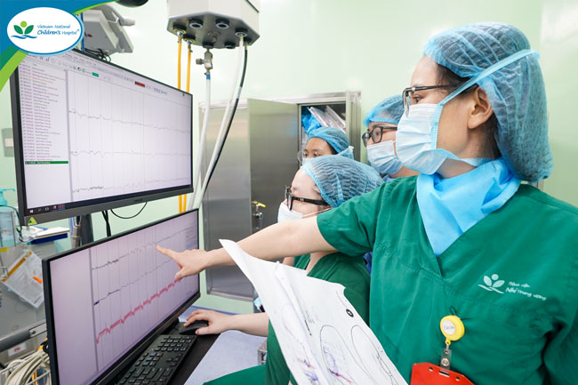
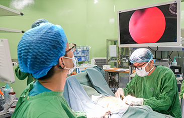
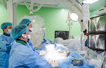
Dr. Nguyen Thi Thao, MSc, MD (Specialist Level II) interpreted EEG findings directly in the operating room to accurately identify the epileptogenic zone and support the surgeons in making the optimal intervention decision.
The greatest challenge in this patient was that the epileptogenic zone was located close to important motor and sensory areas of the brain. Therefore, surgery had to remove the seizure-generating area while preserving neurological function as much as possible.
“We had to calculate the resection margin very carefully. If too little is removed, the child may continue to have seizures; if too much is removed, neurological function may be affected. That is why intracranial EEG and cortical functional mapping before surgery are essential,” Dr. Le Nam Thang shared.
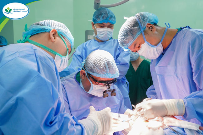
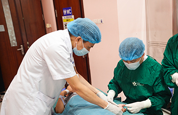
Dr. Le Nam Thang and the surgical team performed the operation for the patient.
Currently, 30 days after surgery, the patient has been completely free of seizures. Her health is progressing well, and she is gradually reintegrating into daily life after many years living with epilepsy.
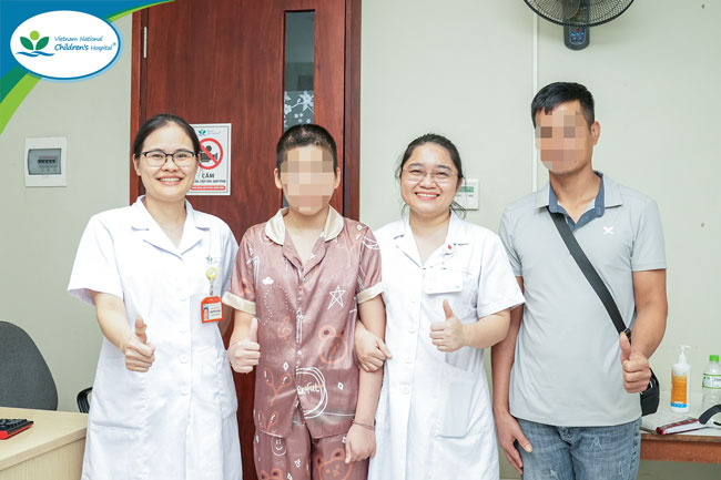
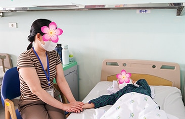
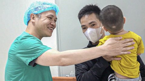
Joy returned as the patient no longer had seizures after surgery.
Mastering epilepsy surgery techniques to international standards
Epilepsy surgery is a highly specialized field in pediatric neurology, requiring close coordination among multiple specialties, including neurology, neurosurgery, diagnostic imaging, anesthesiology and critical care, and others.
Routine application of modern techniques—such as prolonged video-EEG monitoring, brain PET imaging, intracranial EEG, cortical functional mapping, and multidisciplinary consultation—has helped improve the effectiveness of diagnosis and treatment for drug-resistant epilepsy in children.
Dr. Cao Vu Hung, MD, PhD, Director of the Neurology Center, said this was a complex case requiring close coordination among the Division of Neurology, Division of Neurosurgery, and Neurology Functional Exploration Unit to identify the epileptogenic zone. Although MRI showed no lesion, doctors were still able to accurately locate the seizure focus and successfully perform surgery.
This result highlights the importance of meticulous clinical examination combined with advanced diagnostic techniques and specialized knowledge of functional brain anatomy and epilepsy networks.
This case demonstrates the maturity and national pioneering role of the Epilepsy Surgery Group at the Neurology Center, Vietnam National Children’s Hospital, in implementing advanced techniques. It also affirms the Hospital’s growing capacity in diagnosing and treating drug-resistant epilepsy, contributing to improved quality of care and treatment for children with epilepsy in Viet Nam.
Digital Information Office – Training and Research Institute for Child Health