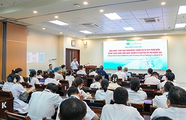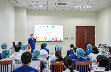
On 11 August 2026, Viet Nam National Children’s Hospital (VNCH) organized a scientific seminar entitled “Pediatric Vasculitis: Recognition and Management from an Imaging Perspective,” featuring Prof. Manphool Singhal, an imaging specialist from the Postgraduate Institute of Medical Education and Research (PGIMER), India.
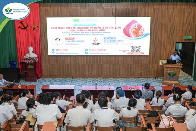
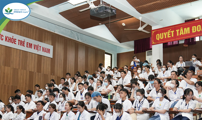
Nearly 400 participants and healthcare professionals from hospitals across the country joined the scientific seminar, both onsite and online, to update their professional knowledge.
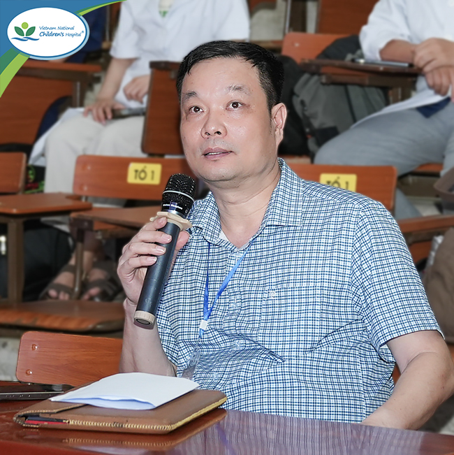
The seminar was chaired by Dr. Tran Phan Ninh, MD, PhD – Head of the Department of Radiology, Viet Nam National Children’s Hospital.
During the seminar, Prof. Manphool Singhal provided updates on approaches to vasculitis based on the size of the affected blood vessels, while emphasizing the increasingly important role of medical imaging in detecting vascular involvement, assessing the extent of lesions, and monitoring disease progression.
Pediatric vasculitis encompasses a diverse group of diseases that can affect multiple organ systems. Depending on the size of the affected vessels, various imaging modalities may be selected for diagnosis, including ultrasound, echocardiography, computed tomography (CT), magnetic resonance imaging (MRI), and positron emission tomography (PET) – an imaging technique that can identify areas of active inflammation and lesions within the body. These advanced imaging modalities not only enable assessment of the vascular lumen but also provide valuable information on the vessel wall, perivascular tissues, target organs, and extravascular involvement.
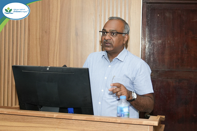
Prof. Manphool Singhal shared in-depth updates on the role of medical imaging in the detection and management of pediatric vasculitis.
The lecture highlighted the shift from invasive procedures toward non-invasive imaging modalities. In medium-vessel vasculitis, multidetector computed tomography angiography (MDCT angiography) and MRI can identify arterial abnormalities as well as target-organ involvement. In large-vessel vasculitis, CT, MRI, and PET-CT/PET-MRI can help determine the extent of vascular involvement, assess disease activity, and monitor disease progression over time. Meanwhile, imaging modalities also play an important role in the assessment of small-vessel vasculitis, depending on the clinical manifestations and organs involved.
Prof. Manphool Singhal illustrated the role of medical imaging through representative clinical cases involving conditions such as polyarteritis nodosa (PAN), Kawasaki disease, deficiency of adenosine deaminase 2 (DADA2), granulomatosis with polyangiitis (GPA), eosinophilic granulomatosis with polyangiitis (EGPA), IgA vasculitis, and Behçet’s disease, a systemic inflammatory vasculitic disorder with diverse manifestations involving the oral cavity, eyes, skin, and blood vessels.
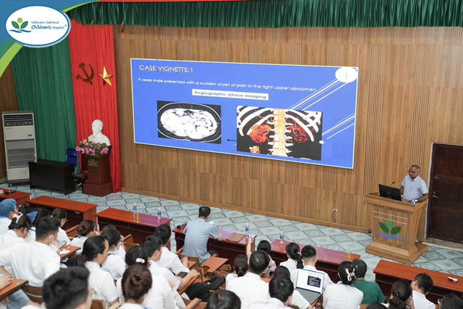
Real-world clinical cases provided participants with a more practical and visual understanding of the role of medical imaging in assessing vasculitic involvement in children.
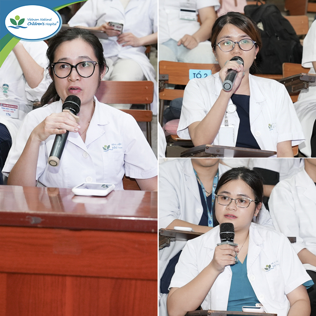
The discussion session was highly interactive, with extensive professional exchange between healthcare professionals from Viet Nam National Children’s Hospital and the international expert.
The scientific seminar reaffirmed the important role of multimodality imaging throughout the management of vasculitis, from diagnosis and assessment of disease severity and extent to long-term follow-up. It also provided an opportunity for healthcare professionals at Viet Nam National Children’s Hospital to engage directly with an international expert, update their knowledge of advances in medical imaging, and strengthen professional collaboration, thereby contributing to enhanced capacity for the detection, assessment, and management of pediatric vasculitis.